Image-based prediction of tumor growth
Participants:
Thierry Colin, Olivier Saut, Julie Joie, Patricio Andrés
Cumsille Atala, Julien Jouganous, Guillaume Lefèvre.
Clinicians involved:
J. Palussière, F. Cornelis, G. Kantor, H. Loiseau and H.
Fathallah .
The idea of this project is to predict the time
development of a clinical tumor or the response to a
treatment using nonlinear PDE models and a series of
CT-Scan or MRI of a patient as inputs. We start by
extracting from our large model a "simple" PDE model
involving few parameters (let's say around 5 independent
parameters). Then we try to find the "best" values of the
parameters that allow to match with the series of image by
solving an optimization problem; then me make a prediction
using this set of parameters. We have tested this strategy
on metastasis to the lung from distant tumors (kidney,
bladder, thyroid). We dispose of time series of CT-scans
of patients that are only under monitoring (i.e. without
treatment). We use only two CT-scans to parametrize our
problem and try to recover the following ones.
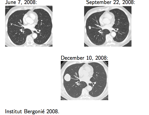
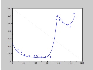
Lung metastasis
The test case presented below concerns a metastasis to the
lung of a bladder tumor. On the left are the 3 CT-scans
that constitute our data set. No treatment was given to
the patient during this period. We used only the image of
June and September to perform the simulation. In the
middle plot, measured volumes of the metastasis (based on
the scan) in time are the circle points while the
continuous line is the volume predicted by the simulation.
On the right images, the tumor given by the simulation for
September and December is in red (top row). Other examples
for metastasis to the lung are given in the publications.
T. Colin, A. Iollo, D. Lombardi, O. Saut System
Identification in Tumor Growth Modeling Using
Semi-empirical Eigenfunctions. Math. Models Methods Appl.
Sci. 22, 1250003 (2012).
T. Colin, A. Iollo, D. Lombardi and O. Saut, Prediction of
the Evolution of Thyroidal Lung Nodules Using a
Mathematical Model, ERCIM News, No 82, pp. 37-38, July
2010.
 |
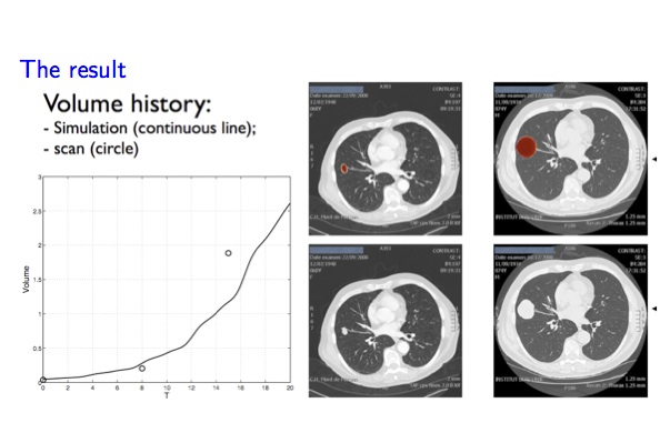 |
Lung metastasis growth prediction. Left: Data. Right:
Model simulations and predictions.
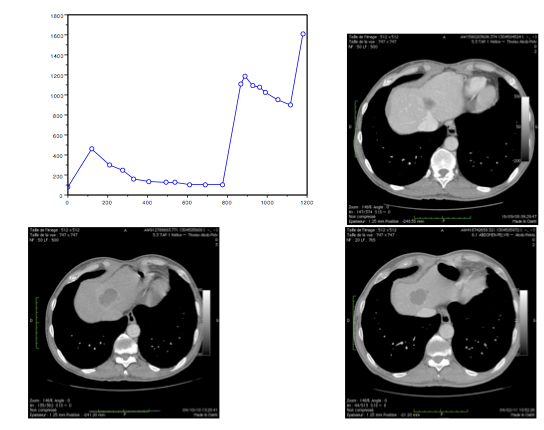
Liver metastasis
We have also some preliminary results concerning
metastasis to the lung of a GIST (Gastro-Intestinal
Stromal Tumor). When the metastasis is discovered, the
patient receives a targeted therapy called Imatinib until
he escapes the treatment. He then receives a second
therapy with Sunitinib until the next escape time. In the
example below we show on the left the clinical data: time
curve of the volume of the metastasis measured on the
successive CT-scans and the CT-scans corresponding (from
left to right and from top to bottom) to the control by
the first treatment, then the escape and then the control
but the second treatment. Right images is a fit of the
model to the volume data.
 |
 |
Liver metastasis. Left: Data provided by the Institut
Bergonié. Right: Model fit.
Glioblastoma (GBM)
With H. Fathallah (University of Alabama at Birmingham),
we have developed a 3D model of glioblastoma expansion
that shows the three layer structure of GBM: a necrotic
core, a proliferative rim surrounded by a cloud of
invasive cells. These elements can be seen on different
sequences of MRI (T1, T1 gado and Flair). It is still an
open problem to characterize precisely these elements on
these images. Below is shown a simulation of GBM expansion
together with an MRI and a biopsy of a glioblastoma.
Th. Colin, A. Iollo, J.-B. Lagaert and O. Saut, An inverse
problem for the the recovery of the vascularization of a
tumor.
Th. Colin, H. Fathallah, J.-B. Lagaert, O. Saut, A
Multilayer Model for GBM: Effects of Invasive Cells and
Anti-Angiogenesis on Growth. Submitted.
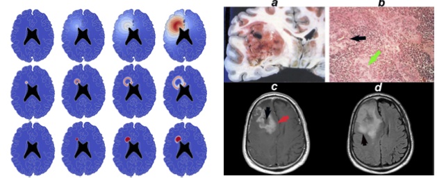
Glioblastoma. Left: Model simulations. Right: Data
provided by the University of Alabama.