Cancer modeling
Participants: Angelo Iollo, Thierry
Colin, Clair Poignard, Olivier Saut.
As in microfluidics, the growth of a tumor is a low Reynolds number flow. Several kinds of interfaces are present (membranes, several populations of cells,...) The biological nature of the tissues impose the use of different models in order to describe the evolution of tumor growth. The complexity of the geometry, of the rheological properties and the coupling with multi-scale phenomena is high but not far away from those encountered in microfluidics and the models and methods are close. The challenge is twofold. On one hand, we wish to understand the complexity of the coupling effects between the different levels (cellular, genetic, organs, membranes, molecular). Trying to be exhaustive is of course hopeless, however it is possible numerically to isolate some parts of the evolution in order to better understand the interactions. Another strategy is to test in silico some therapeutic innovations.
It is therefore useful to model a tumor growth at several stage of evolution. The macroscopic continuous model is based on Darcy’s law which seems to be a good approximation to describe the flow of the tumor cells in the extra-cellular matrix. It is therefore possible to develop a two-dimensional model for the evolution of the cell densities. We formulate mathematically the evolution of the cell densities in the tissue as advection equations for a set of unknowns representing the density of cells with position (x; y) at time t in a given cycle phase. Assuming that all cells move with the same velocity given by Darcy’s law and applying the principle of mass balance, one obtains the advection equations with a source term given by a cellular automaton. We assume diffusion for the oxygen and the diffusion constant depends on the density of the cells. The source of oxygen corresponds to the spatial location of blood vessels. The available quantities of oxygen interact with the proliferation rate given by the cellular automaton.
A forthcoming investigation in cancer treatment simulation is the influence of the electrochemotherapy on the tumor growth. Electrochemotherapy consists in imposing to the malignant tumor high voltage electric pulses so that the plasma membrane of carcinoma cells is permeabilized. Biologically active molecules such as bleomycin, which usually cannot diffuse through the membrane, may then be internalized. A work in progress consists in modelling electromagnetic phenomena at the cell scale. A coupling between the microscopic description of the electroporation of cells and its influence on the global tumor growth at the macroscopic scale is expected. Another key point is the parametrization of the models in order to produce image-based simulations.
The second challenge is more ambitious. Mathematical models of cancer have been extensively developed with the aim of understanding and predicting tumor growth and the effects of treatments. In vivo modeling of tumors is limited by the amount of information available. However, in the last few years there have been dramatic increases in the range and quality of information available from non-invasive imaging methods, so that several potentially valuable imaging measurements are now available to quantitatively measure tumor growth, assess tumor status as well as anatomical or functional details. Using different methods such as the CT scan, magnetic resonance imaging (MRI), or positron emission tomography (PET), it is now possible to evaluate and define tumor status at different levels: physiological, molecular and cellular. In this context, the present project aims at supporting the decision process of oncologists in the definition of therapeutic protocols via quantitative methods. The idea is to build mathematically and physically sound phenomenological models that can lead to patient-specific full-scale simulations, starting from data collected typically through medical imagery like CT scans, MRIs and PET scans or by quantitative molecular biology for leukemia. Our ambition is to provide medical doctors with patient-specific tumor growth models able to estimate, on the basis of previously collected data and within the limits of phenomenological models, the evolution at subsequent times of the pathology and possibly the response to the therapies. The final goal is to provide numerical tools in order to help to answer to the crucial questions for a clinician:
When to start a treatment?
When to change a treatment?
When to stop a treatment?
Also we intend to incorporate real-time model information for improving the precision and effectiveness of non-invasive or micro-invasive tumor ablation techniques like acoustic hyperthermia, electroporation, radiofrequency or cryo-ablation. We will specifically focus on the following pathologies: Lung and liver metastasis of a distant tumor Low grade and high grade gliomas, meningiomas Chronic myelogenous leukemia. These pathologies have been chosen because of the existing collaborations between the applied mathematics department of University of Bordeaux and the Institut Bergonié.
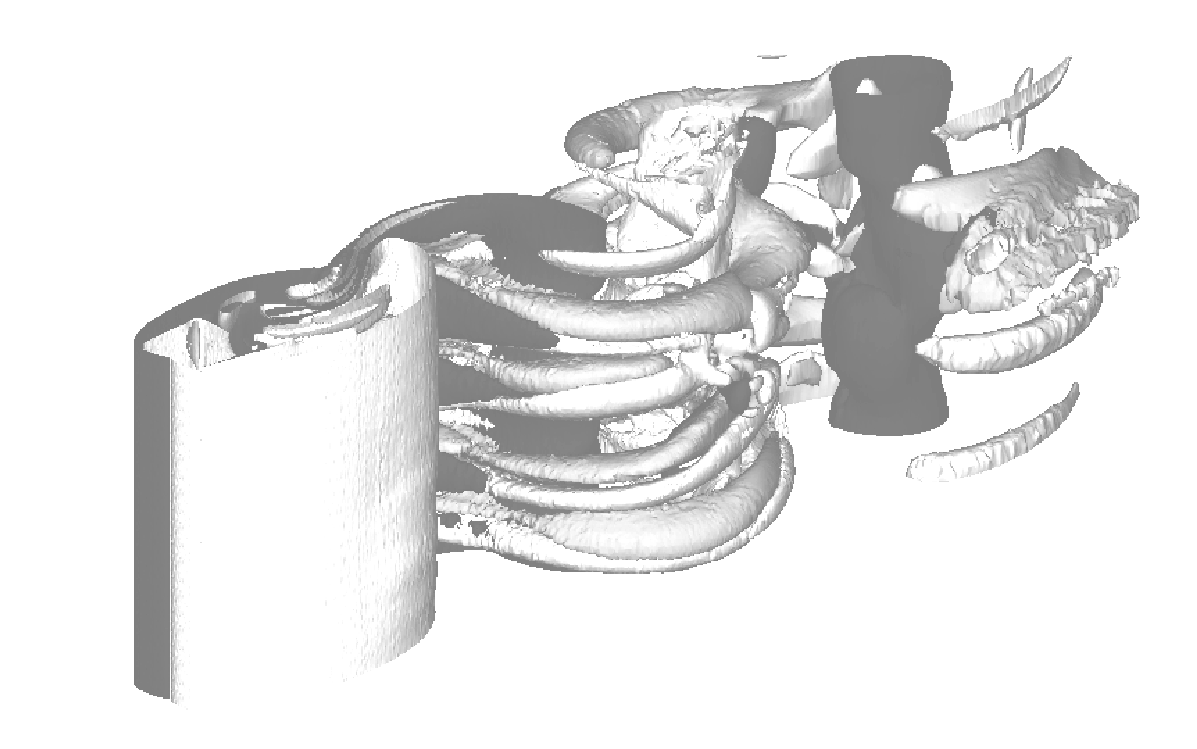
Our approach is deterministic and spatial: it is based on solving an inverse problem based on imaging data. Models are of partial differential equation (PDE) type. They are coupled with a process of data assimilation based on imaging.We already have undertaken test cases on patients that are followed at Bergonié for lung metastases of thyroid tumors. These patients have a slowly evolving, asymptomatic metastatic disease, monitored by CT scans. On two thoracic images relative to successive times, the volume of the tumor under investigation is extracted by segmentation. To test our method, we chose patients without treatment and for whom we had at least three successive
As in microfluidics, the growth of a tumor is a low Reynolds number flow. Several kinds of interfaces are present (membranes, several populations of cells,...) The biological nature of the tissues impose the use of different models in order to describe the evolution of tumor growth. The complexity of the geometry, of the rheological properties and the coupling with multi-scale phenomena is high but not far away from those encountered in microfluidics and the models and methods are close. The challenge is twofold. On one hand, we wish to understand the complexity of the coupling effects between the different levels (cellular, genetic, organs, membranes, molecular). Trying to be exhaustive is of course hopeless, however it is possible numerically to isolate some parts of the evolution in order to better understand the interactions. Another strategy is to test in silico some therapeutic innovations.
It is therefore useful to model a tumor growth at several stage of evolution. The macroscopic continuous model is based on Darcy’s law which seems to be a good approximation to describe the flow of the tumor cells in the extra-cellular matrix. It is therefore possible to develop a two-dimensional model for the evolution of the cell densities. We formulate mathematically the evolution of the cell densities in the tissue as advection equations for a set of unknowns representing the density of cells with position (x; y) at time t in a given cycle phase. Assuming that all cells move with the same velocity given by Darcy’s law and applying the principle of mass balance, one obtains the advection equations with a source term given by a cellular automaton. We assume diffusion for the oxygen and the diffusion constant depends on the density of the cells. The source of oxygen corresponds to the spatial location of blood vessels. The available quantities of oxygen interact with the proliferation rate given by the cellular automaton.
A forthcoming investigation in cancer treatment simulation is the influence of the electrochemotherapy on the tumor growth. Electrochemotherapy consists in imposing to the malignant tumor high voltage electric pulses so that the plasma membrane of carcinoma cells is permeabilized. Biologically active molecules such as bleomycin, which usually cannot diffuse through the membrane, may then be internalized. A work in progress consists in modelling electromagnetic phenomena at the cell scale. A coupling between the microscopic description of the electroporation of cells and its influence on the global tumor growth at the macroscopic scale is expected. Another key point is the parametrization of the models in order to produce image-based simulations.
The second challenge is more ambitious. Mathematical models of cancer have been extensively developed with the aim of understanding and predicting tumor growth and the effects of treatments. In vivo modeling of tumors is limited by the amount of information available. However, in the last few years there have been dramatic increases in the range and quality of information available from non-invasive imaging methods, so that several potentially valuable imaging measurements are now available to quantitatively measure tumor growth, assess tumor status as well as anatomical or functional details. Using different methods such as the CT scan, magnetic resonance imaging (MRI), or positron emission tomography (PET), it is now possible to evaluate and define tumor status at different levels: physiological, molecular and cellular. In this context, the present project aims at supporting the decision process of oncologists in the definition of therapeutic protocols via quantitative methods. The idea is to build mathematically and physically sound phenomenological models that can lead to patient-specific full-scale simulations, starting from data collected typically through medical imagery like CT scans, MRIs and PET scans or by quantitative molecular biology for leukemia. Our ambition is to provide medical doctors with patient-specific tumor growth models able to estimate, on the basis of previously collected data and within the limits of phenomenological models, the evolution at subsequent times of the pathology and possibly the response to the therapies. The final goal is to provide numerical tools in order to help to answer to the crucial questions for a clinician:
When to start a treatment?
When to change a treatment?
When to stop a treatment?
Also we intend to incorporate real-time model information for improving the precision and effectiveness of non-invasive or micro-invasive tumor ablation techniques like acoustic hyperthermia, electroporation, radiofrequency or cryo-ablation. We will specifically focus on the following pathologies: Lung and liver metastasis of a distant tumor Low grade and high grade gliomas, meningiomas Chronic myelogenous leukemia. These pathologies have been chosen because of the existing collaborations between the applied mathematics department of University of Bordeaux and the Institut Bergonié.
Our approach is deterministic and spatial: it is based on solving an inverse problem based on imaging data. Models are of partial differential equation (PDE) type. They are coupled with a process of data assimilation based on imaging.We already have undertaken test cases on patients that are followed at Bergonié for lung metastases of thyroid tumors. These patients have a slowly evolving, asymptomatic metastatic disease, monitored by CT scans. On two thoracic images relative to successive times, the volume of the tumor under investigation is extracted by segmentation. To test our method, we chose patients without treatment and for whom we had at least three successive
Examples: Test